GrabCAD
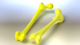
Femur Bone
by GrabCAD
Last crawled date: 1 year, 10 months ago
Femur Bone (with main whole or cavity)
Prepared by Digital Imaging (DI) (CMM Optical) and CT scan (CT Imaging)
Human specimen’s properties for these femoral bones are:
Femur 1: Male, Left leg, Age 44, Death 2016, Weigh 85 (kg), Height 185 (cm) (original scale)
Femur 2: Male, Left leg, Age 69, Death 2015, Weigh 79 (kg), Height 188 (cm) (original scale)
To model the bone in FE software, the Femur 1 and 2 were subjected to the 3D digital imaging (CMM optical 2015, made by GOM Co., Germany with VDA calibration). The digitized model was first modeled as a shell using the cloud points, (i.e. STEP format, in CATIA V5/R21) and then converted to a solid model. It should be mentioned that in this study, the trabecular or marrow bones are not included in finite element model. To model the central cavity and inner layer of the bone, a precise method using the CT scan data is used (Spiral CT scan, ID.NO: ASMA 951355-3801, modern AZMA Co. Siemens, SOMATOM plus4). In this method, the bone is fully scanned and then an array of sections with 10 mm distances with respect to each other are created using the Marco PACS software. The absolute distance of each section from the top of the Femur is used to define the exact coordinate of the sections. All sections are stored in two-dimensional with digital imaging in communication in medicine (DICOM) format and are visualized in Adobe Photoshop CC. Finally, the three-dimensional model of the original Femur along with the 2D images of cross sections are transferred into SolidWorks 2013 software using the exact coordinates of cross sections and the cavity of the bone is modeled using the lofted cut module of the software. Using this method the bone is modeled completely precision of 0.1 mm. Since in this article the critical buckling load of the bone shaft is studied, the proximal and distal regions of Femur are excluded from the model.
Similar models
grabcad
free

Femur Bone
...central hole were determined in separate plans as sketched. similary, all the cross sections were modeled and in the...
grabcad
free

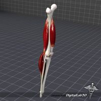
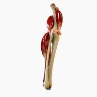
Knee Joint
...y gom, germany with vda calibration), by mimics, solidworks and catia. solid model (noshell) without ligaments . stp/step format.
thingiverse
free

Skull CT Scan w/ Brain Cavity
...skull ct scan w/ brain cavity
thingiverse
this model is taken from a ct scan of a middle aged male, and includes a brain cavity.
grabcad
free

Femur_3D_Model
...femur_3d_model
grabcad
human femur bone 3d model, reconstructed from ct images, modeled by mimics software.
grabcad
free

Human Femur
...
the dicom image set was obtained from www.embodi3d.com. it is named "samapple_ct1" uploaded by the user netlimit2000.
thingiverse
free

Broken Femur / Bone Screw Model Prop
... so that the models would fit on the printer. this includes the models for the bone-screws, which were also printed at 80% scale.
thingiverse
free

Optical CT Scan parts by EmyT
...n-and-photogrammetry/
these parts were made using freecad. you can open the .fcstd files with this free and open-source software.
grabcad
free

Human Tibia Bone
...
human specimen (male). optical cmm modelling (made by gom, germany with vda calibration), by solidworks. solid model (noshell).
grabcad
free

Human Fibula Bone
...
human specimen (male). optical cmm modelling (made by gom, germany with vda calibration), by solidworks. solid model (noshell).
thingiverse
free

CT Scan of Mouse Femur (half) by 10monthspregnant
...ct scan of mouse femur (half) by 10monthspregnant
thingiverse
microct scan of a mouse femur
Femur
turbosquid
$29

Human Femur
... available on turbo squid, the world's leading provider of digital 3d models for visualization, films, television, and games.
turbosquid
$149

Spiral Fracture skeleton labelled femur
...spiral fracture skeleton labelled femur for download as blend on turbosquid: 3d models for games, architecture, videos. (1480589)
3d_export
$44
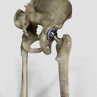
Hip Replacement
...that is placed into the hollow center of the femur ...
3d_export
$35
Skeleton 3D Model
...man skeleton skeletal system bone skull spine vertabrae ribcage femur jaw teeth anatomy mandible death body character rib skeleton...
3d_export
$5

Bone axe
...axe made of bones. it was inspired by a femur of a dead deer i saw in the forest....
cg_studio
$45
Human skeleton3d model
...human skeletal system human skeleton bones bone spine skull femur teeth skeleton system .obj .mb .max .ma .fbx .dae...
3d_export
$179
Knee Joint Anatomy 3D Model
...model 3dexport digitallab3d human knee joint anatomy thigh tibia femur patella largest complicated body people artery leg character ligament...
cg_studio
$179
Knee Joint Anatomy3d model
...model cgstudio digitallab3d human knee joint anatomy thigh tibia femur patella largest complicated body people artery leg character ligament...
3d_export
$5

tibia implant real model for training
...collar comes into contact with the sawdust of the femur in places where the implant comes into contact with...
3d_export
$34

Knee Replacement
...during a total knee replacement, the end of the femur bone is removed and replaced with a metal...
Bone
3d_export
$5

Bone
...bone
3dexport
bone
3d_export
$5

bone
...bone
3dexport
geological fossil/ extinct dinosaur's bone
3ddd
$1

Roshe | Bone
...roshe | bone
3ddd
bone , roshe
bone диван
3ddd
$1

Roshe | Bone
...| bone
3ddd
bone , roshe , угловой
bone угловой
3ddd
$1

Roshe | Bone
...roshe | bone
3ddd
bone , roshe
кресло и пуф bone
turbosquid
$2

Bone
...
royalty free 3d model bone for download as ma, obj, and fbx on turbosquid: 3d models for games, architecture, videos. (1254908)
3ddd
$1

bone chair
...bone chair
3ddd
joris laarman , bone chair
стул bone chair от joris laarman
turbosquid
$29

Bone
... available on turbo squid, the world's leading provider of digital 3d models for visualization, films, television, and games.
turbosquid
$25

Bone
... available on turbo squid, the world's leading provider of digital 3d models for visualization, films, television, and games.
turbosquid
$7

Bone
...ree 3d model bone for download as 3ds, obj, fbx, dae, and stl on turbosquid: 3d models for games, architecture, videos. (1442970)