Thingiverse

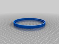
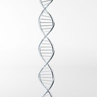
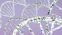
Photo 51: x-ray diffraction pattern of DNA
by Thingiverse
Last crawled date: 4 years, 4 months ago
Original image file: https://en.wikipedia.org/wiki/File:Photo_51_x-ray_diffraction_image.jpg
Converted to lithophane using: http://3dp.rocks/lithophane/
From Wikipedia:
Photo 51 is the name given to an X-ray diffraction image of a paracristalline gel composed of DNA fiber (often misinterpreted as crystallized DNA) taken by Raymond Gosling, a graduate student working under the supervision of Rosalind Franklin in May 1952 at King's College London, while working in Sir John Randall's group.
James Watson was shown the photo by his collaborator, Maurice Wilkins, without Rosalind Franklin's knowledge.
Wilkins did this, as by this time, Gosling had returned under his supervision, since Franklin was leaving King's and Randall had asked Gosling to share all his data with Wilkins. Along with Francis Crick, Watson used characteristics and features of Photo 51, together with evidence from multiple other sources, to develop the chemical model of the DNA molecule. Their model, and manuscripts by Wilkins and colleagues, and Gosling and Franklin, were first published, together, in 1953, in the same issue of Nature. In 1962, the Nobel Prize in Physiology or Medicine was awarded to Watson, Crick and Wilkins.
The prize was not awarded to Franklin; she had died four years earlier, and although there was not yet a rule against posthumous awards, the Nobel Committee generally does not make posthumous nominations. Likewise, Gosling's work was not cited by the prize committee.
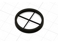
Similar models
blendswap
free

Fake DNA X-ray Diffraction by Rosalind Franklin
...klin in cycle.
spherical roughness of glass and sobel filter make the blur effect.
the node setting and behind the scene ishere.
3dwarehouse
free

Himingway House in a Box
...ctions, and three nonfiction works were published posthumously. many of his works are considered classics of american literature.
3dwarehouse
free

ANDi's_Hall
...i's_hall
3dwarehouse
andi's hall, that was built for andi's awards. #andi #andis #award #awards #hall #prize #prizes
3dwarehouse
free

Polartic (Rosalind Franklin)
... franklin (ex finnclipper di finnlines - grimaldi lines group) di balearia, in livrea di ventouris ferries (nuovo nome: polartic)
thingiverse
free

Toni Morrison Lithophane
...t, then imported it as a part to their standard cad environment for fine-tuning with the lithophane.
https://youtu.be/8d_rtm-jnp0
turbosquid
$29

Watson Table DNA
... available on turbo squid, the world's leading provider of digital 3d models for visualization, films, television, and games.
3dwarehouse
free

Nobel Peace Center
... in oslo in 1872, it has been changed and it's now the famous nobel peace center. #nobel #oslo #peace #prize #vestbanen #vika
thingiverse
free

Penrose Trivet by Penguinmd
...et made from penrose tiling. roger penrose won the nobel prize yesterday for his work on black holes. seems fitting to celebrate.
cg_trader
$4

Chess King
...he chess table with a wood texture. chess gameplan victory win competition statue sculpture award medal prize triumph sports game
thingiverse
free

Alnico quasi chrystal pattern by kokr
...spired by an illustration of the quasi chrystal alnico pattern published by the nobel committee for the work of daniel schechtman
Diffraction
thingiverse
free
removable diffraction spike mask by K_Bahr
...removable diffraction spike mask by k_bahr
thingiverse
removable diffraction spike mask
thingiverse
free

Frame for Diffraction Grating by owensscience
... two of these, and glued the diffraction film in between them to prevent my students from getting fingerprints all over the film.
thingiverse
free

Stand for linear diffraction grating by antiElectron
... grating by antielectron
thingiverse
this is a simple stand for linear diffraction grating, ideal for experimenting with lasers.
thingiverse
free

Removable Diffraction Spike Mask by K_Bahr
...sk by k_bahr
thingiverse
the size should fit on telescopes with 120mm to 127mm
aperture (5inch)
removable diffraction spike mask
thingiverse
free
Diffraction Spike Mask Skywatcher Esprit 80ED by castropic
...diffraction spike mask skywatcher esprit 80ed by castropic
thingiverse
diffraction spike mask for skywatcher esprit 80ed
thingiverse
free

Laser Diffraction Grating by jsteuben
...e wave/particle duality of light. see http://en.wikipedia.org/wiki/double-slit_experiment for further details on this experiment.
thingiverse
free

Laser Diffraction Pattern Cap
...ous size laser pointers.
more detailhttps://www.olympus-lifescience.com/en/microscope-resource/primer/lightandcolor/interference/
blendswap
free

Gem Node Diffraction
...s not work in all types of light so be careful. you can easily turn up the color effect in the compositor or contact me for help.
thingiverse
free

X-Ray diffraction sample piece by BenWittbrodt
...nto the sample holder of a scintag xds-2000 powder diffractometer without any sample prep. corners rounded for ease of printing.
thingiverse
free
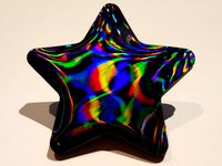
Star for Diffraction Grating Test
...ext! the colors look best under strong, direct light. very cool!
more of my models on cults:https://cults3d.com/en/users/abbymath
Dna
3d_export
$19

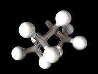
DNA
...dna
3dexport
3d model of the dna textures 8k
design_connected
$9
DNA
...dna
designconnected
next home collection dna computer generated 3d model. designed by hopf, benjamin.
3d_export
$5

DNA
...dna
3dexport
3d_ocean
$1
DNA Strand
...dna strand
3docean
d.n.a dna strand
accurate 3d model of a dna strand
turbosquid
free

DNA
...dna
turbosquid
free 3d model dna for download as max on turbosquid: 3d models for games, architecture, videos. (1497359)
turbosquid
$9
DNA
...a
turbosquid
royalty free 3d model dna for download as blend on turbosquid: 3d models for games, architecture, videos. (1667313)
turbosquid
$1

Dna
...dna
turbosquid
royalty free 3d model dna for download as fbx on turbosquid: 3d models for games, architecture, videos. (1410365)
3ddd
$1
Next / DNA
...next / dna
3ddd
next , dna
http://www.next.de/cms/en/dna-en
turbosquid
$45

DNA
...
royalty free 3d model dna for download as max, fbx, and obj on turbosquid: 3d models for games, architecture, videos. (1622745)
turbosquid
free

DNA
...
free 3d model dna for download as ma, c4d, lwo, max, and obj on turbosquid: 3d models for games, architecture, videos. (1521540)
51
3ddd
$1
Pillows 51
...pillows 51
3ddd
подушка
pillows 51
turbosquid
$20

51
... available on turbo squid, the world's leading provider of digital 3d models for visualization, films, television, and games.
3d_export
$18

plants 51
...plants 51
3dexport
plants 51 is a collection of flower plants in both hanging pot and flower stand,, hope you like it.
turbosquid
$10

Bedcloth 51
...osquid
royalty free 3d model bedcloth 51 for download as max on turbosquid: 3d models for games, architecture, videos. (1522737)
turbosquid
$15

Curtain 51
...
royalty free 3d model curtain 51 for download as max and obj on turbosquid: 3d models for games, architecture, videos. (1350126)
turbosquid
$15

Chair 51
...alty free 3d model chair 51 for download as max, obj, and fbx on turbosquid: 3d models for games, architecture, videos. (1497282)
turbosquid
$15

Sofa 51
...yalty free 3d model sofa 51 for download as max, obj, and fbx on turbosquid: 3d models for games, architecture, videos. (1503071)
turbosquid
$10

Lamp 51
...yalty free 3d model lamp 51 for download as max, obj, and fbx on turbosquid: 3d models for games, architecture, videos. (1500778)
turbosquid
$6

Table 51
...alty free 3d model table 51 for download as max, obj, and fbx on turbosquid: 3d models for games, architecture, videos. (1503874)
turbosquid
$39
Ka-51
... available on turbo squid, the world's leading provider of digital 3d models for visualization, films, television, and games.
Pattern
3d_export
$10
pattern
...pattern
3dexport
old carved pattern
3d_ocean
$4

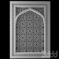
Window Pattern
...c4d cinema4d design element geometry geometry pattern grill pattern pattern vray vrayforc4d window pattern
window or wall pattern
design_connected
$18
Pattern
...pattern
designconnected
emu group pattern computer generated 3d model. designed by levy, arik.
3ddd
$1
pattern
...pattern
3ddd
решетка
1000x1000
3ddd
$1

Stucco pattern
...stucco pattern
3ddd
розетка
stucco pattern
3d_ocean
$10

Pattern
...pattern
3docean
extrude
national ornament
turbosquid
$6

Pattern
... available on turbo squid, the world's leading provider of digital 3d models for visualization, films, television, and games.
3d_export
$5

pattern
...pattern
3dexport
cnc router --3d printer
turbosquid
$25

Wall Pattern A05 (pattern only)
... available on turbo squid, the world's leading provider of digital 3d models for visualization, films, television, and games.
turbosquid
$25
Wall Pattern A04 (pattern only)
... available on turbo squid, the world's leading provider of digital 3d models for visualization, films, television, and games.
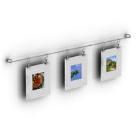
Photo
3ddd
$1
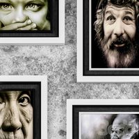
photo frame
...photo frame
3ddd
photo frame
3ddd
$1

Photo Frame
...photo frame
3ddd
рисунок
photo frame
3d_export
$5

photo frame
...or photos, pictures. you can put shes anywhere. in the kitchen, on the table in the bedroom, on the shelf in the hall, and so on.
archive3d
free
Photos 3D Model
...tos photo photos set
set photos n160216 - 3d model (*.gsm+*.3ds) for interior 3d visualization.
3d_export
$5
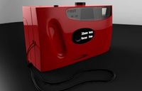
Photo 3D Model
...photo 3d model
3dexport
photo
photo 3d model max140588 62242 3dexport
3d_export
$14

photo printer
...g projects, and was originally modeled in 3ds max 2012 and rendered with v-ray. renders have no postprocessing. hope you like it!
turbosquid
$10

photo frame
...osquid
royalty free 3d model photo frame for download as obj on turbosquid: 3d models for games, architecture, videos. (1404417)
turbosquid
$9

Photo studio
...uid
royalty free 3d model photo studio for download as blend on turbosquid: 3d models for games, architecture, videos. (1498830)
turbosquid
$5

Photo Frame
...royalty free 3d model photo frame for download as fbx and upk on turbosquid: 3d models for games, architecture, videos. (1163533)
turbosquid
$2

Photo Frame
...royalty free 3d model photo frame for download as max and fbx on turbosquid: 3d models for games, architecture, videos. (1352878)
Ray
design_connected
$16
Ray
...ray
designconnected
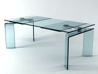
fiam italia ray dining tables computer generated 3d model. designed by bartoli design.
turbosquid
$5
Rays
...
royalty free 3d model rays for download as max, obj, and fbx on turbosquid: 3d models for games, architecture, videos. (1706935)
3ddd
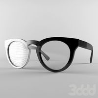
$1
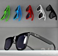
ray ban
...ray ban
3ddd
ray ban
ray ban
3ddd
$1
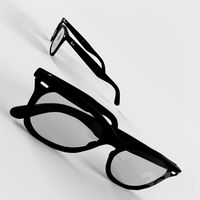
Ray Ban
...ray ban
3ddd
ray ban , очки
очки ray ban
3d_ocean
$15
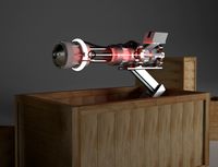
Ray Gun
...gun shrink ray
who wouldn’t want a ray gun in a box. all objects are editable and able to be textured. textures are not included.
3ddd
$1
Ray Ban
...ray ban
3ddd
очки
очки ray ban
3ddd
$1
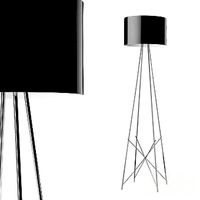
Торшер Ray F. Ray F. Италия
...торшер ray f. ray f. италия
3ddd
flos
торшер ray f. ray f. италия 1280х360
3ddd
$1
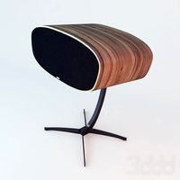
davone ray
...y
3ddd
колонка , davon , ray
davone_ray h 734
design_connected
$18
Sting Ray
...sting ray
designconnected
tuuci sting ray computer generated 3d model.
design_connected
$13
Ray-S
...ray-s
designconnected
ray-s computer generated 3d model. designed by dordoni, rodolfo.