GrabCAD

Femur Bone
by GrabCAD
Last crawled date: 1 year, 10 months ago
Femur Bone
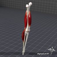
Prepared by Digital Imaging (DI) (CMM Optical) and CT Scan (CT Imaging)
Human specimen’s properties for this Femur bone are: Male, Left leg, Age 44, Death 2016, Weigh 85 (kg), Height 185 (cm) (original scale)
Femur bone was created in 3D using Digital Imaging (DI) (CMM Optical 2015, Made by GOM Co., Germany). Digitized model was first modeled as Shell and cloud points, i.e. STEP format, in CATIA V5/R21 and then was surfaced as a Solid. The inner area of the Femur (the central cavity and inner layer) cannot be modeled and surfaced using this method. So to model this section (which is one of the most important parts in FE model), Computed Tomography (CT) scan method is used (Only the Cortical bone tissue is modeled using this approach and Trabecular tissue is not modeled). In this way, the bone is completely and precisely modeled. In this type of imaging, first, all the bone is scanned and then sections with a space of 10mm of each other and with an exact coordinates from the top of Femur bone (Proximal region) to the end part (Distal region) of the bone is created using the related software. All sections are stored in two-dimensional and with Digital Imaging and Communication in Medicine (DICOM) format (Using Marco PACS and SECTRA PACS or Sonic PACS software and DICOM files, all the sections, i.e. 25 sections in diaphysis region with a distance of 10mm and almost 45 sections in throughout of bone), the output format of CT scanner, and visualized in Photoshop CC. Finally, the three-dimensional model of the original bone along with 2D images of cross sections of CT scan separately with the desired scaling transferred into Solid Works 2013 and each cross section corresponded to the outer boundaries of the same cross section in 3D models so that its central hole were determined in separate plans as sketched. Similarly, all the cross sections were modeled and in the end, using the lofted cut modules (removed) option all the sketches emptied from the original bone volume. This method is accurate to 0.01mm.
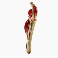
Similar models
grabcad
free

Femur Bone
...le the critical buckling load of the bone shaft is studied, the proximal and distal regions of femur are excluded from the model.
grabcad
free

Human Femur
...
the dicom image set was obtained from www.embodi3d.com. it is named "samapple_ct1" uploaded by the user netlimit2000.
thingiverse
free

Broken Femur / Bone Screw Model Prop
... so that the models would fit on the printer. this includes the models for the bone-screws, which were also printed at 80% scale.
grabcad
free

Knee Joint
...y gom, germany with vda calibration), by mimics, solidworks and catia. solid model (noshell) without ligaments . stp/step format.
cg_trader
$19

Anatomy bone femoral
...anatomy bone femoral
cg trader
3d bone femur (cross-section) .
cg_trader
$15

Human Pelvis | 3D
...human pelvis | 3d
cg trader
antomical human pelvis created using ct scan dicom images.
grabcad
free

Femur_3D_Model
...femur_3d_model
grabcad
human femur bone 3d model, reconstructed from ct images, modeled by mimics software.
cg_trader
$15
Human Lungs | 3D
...human lungs | 3d
cg trader
anatomically accurate human lungs 3d model for 3d printing.
created using ct scan dicom images.
grabcad
free

DICOM to CAD - Torso (Bone)
...d - torso (bone)
grabcad
this is a dicom to cad conversion of a human torso. region of interest is bone. this can be 3d printed.
cg_trader
$15

Mandable from DICOM - | 3D
...ndable from dicom - | 3d
cg trader
3d model created using human ct scan dicom data set which represents accurate human anatomy.
Femur
cg_studio
$45
Human skeleton3d model
...human skeletal system human skeleton bones bone spine skull femur teeth skeleton system .obj .mb .max .ma .fbx .dae...
cg_studio
$99

Human Larynx - Anatomy3d model
...organs stem surgery man face spine vertebra rib ribcage femur reaper fracture dead death head character pirate .3ds .x...
cg_studio
$45
Human Skeleton: Rentgen View RenderReady3d model
...clavicle sternum scapula ribs humerus ulna vertebral pelvic girdle femur .3ds .c4d .dxf .fbx .lwo .max .obj .xsi -...
cg_studio
$70
Human Skeleton in Body Anatomy3d model
...clavicle sternum scapula ribs humerus ulna vertebral pelvic girdle femur .stl .obj .max .fbx .c4d .3ds - human skeleton...
3d_export
$179
Knee Joint Anatomy 3D Model
...model 3dexport digitallab3d human knee joint anatomy thigh tibia femur patella largest complicated body people artery leg character ligament...
cg_studio
$179
Knee Joint Anatomy3d model
...model cgstudio digitallab3d human knee joint anatomy thigh tibia femur patella largest complicated body people artery leg character ligament...
cg_studio
$80
Female Skeleton in transparent Woman Body3d model
...clavicle sternum scapula ribs humerus ulna vertebral pelvic girdle femur .stl .3dm .obj .max .lwo .fbx .c4d .3ds -...
3d_export
$35
Skeleton 3D Model
...man skeleton skeletal system bone skull spine vertabrae ribcage femur jaw teeth anatomy mandible death body character rib skeleton...
3d_export
$44
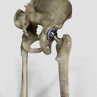
Hip Replacement
...that is placed into the hollow center of the femur ...
3d_export
$5

Bone axe
...axe made of bones. it was inspired by a femur of a dead deer i saw in the forest....
Bone
3d_export
$8

ChiropractorTable 3D Model
...chiropractortable 3d model 3dexport medical doctor bone poser table bed 3d model hospital clinic office chiropractortable...
3d_export
$40
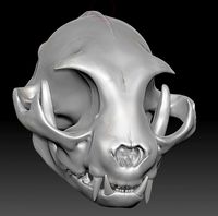
Cat Skull ZBrush Sculpture 3D Model
...skull zbrush sculpture 3d model 3dexport cat feline skull bone kitty cranium pet cat skull zbrush sculpture 3d model...
3d_export
$79

Abraham Simpson rigged 3D Model
...toon serie tv rigged rig animation skin biped character bone abraham simpson rigged 3d model fabelar 28584...
3d_export
$50

Human Spine 3D Model
...human spine 3d model 3dexport spine skeleton vertebrae bone human anatomy body medical cervical thoracic lumbar sacrum coccyx...
3ddd
$1
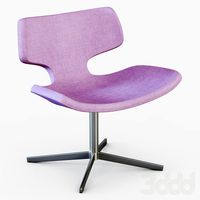
Armchair BONE
...armchair bone
3ddd
materia , bone
3ds max 2010.v-ray 2.40.03.file formats fbx,obj,3ds
3ddd
$1

Oluce Empty 439
...3ddd oluce , empty oluce empty 439, designer jörg bone ø cm. 59 ↓ cm. 32 file contain 3...
3d_ocean
$5

ELEGANT SCULL 3D MODEL
...elegant scull 3d model 3docean abstract anatomy body bone bone dead death die halloween head human jaw lines male...
3d_ocean
$29

Cartoon Skeleton Rigged
...cartoon skeleton rigged 3docean body bone cartoon chararcter funny halloween horror mentalray monster rigged skeleton...
3d_export
$25
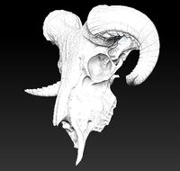
Ram skull 3D Model
...mammal ram horn vray skeletal skeleton skull decor decoration bone head 3dprinter printable ram skull 3d model download .c4d...
cg_studio
$45
Human skeleton3d model
...skeleton3d model cgstudio human skeletal system human skeleton bones bone spine skull femur teeth skeleton system .obj .mb .max...